After a diagnosis, it’s natural to wonder what’s next. This guide will help you understand the tests you may need and why they are important.
A colorectal cancer diagnosis can be overwhelming. Once you have the diagnosis, you’ll likely wonder: Now what?
You’ll have decisions to make with your health care team about your treatment. But first, you’ll likely need more tests. These tests are important; they help your providers understand your cancer better. They need to know how fast it’s growing and if it has spread to other parts of the body.
That information will help your medical care team decide the best way to treat the cancer. This guide will help you learn about the various lab, blood and imaging tests you might have.
Lab tests on biopsy or surgery samples
Colorectal cancer is most often diagnosed by taking out and testing a small piece of a polyp or tumor. This is called a biopsy. The tissue removed is called a sample.
When cancer is found, more tests may be done on the sample. For example, some tests can find genetic changes in the cancer cells. These details about the cancer can help your care team figure out which treatment might be best.
Blood tests
The information provided by different blood tests can be very helpful. These tests are done by taking a small sample of blood.
Complete blood count
A complete blood count (CBC) measures:
- The number of red blood cells
- The number of white blood cells
- The number of platelets
Information from the CBC can help your health care provider know what the cancer is doing to your body. For example, a low red blood cell count (anemia) may be a sign that the tumor is causing bleeding.
The complete blood count is also important during treatment and will be checked often. If you need surgery, your care team will make sure your platelet count is high enough. Having a low platelet count before surgery puts you at greater risk for bleeding.
If chemotherapy is part of your treatment, your provider will check your complete blood count regularly. That’s because chemo can affect your bone marrow, which is where new blood cells are made.
Blood chemistry tests
Blood tests can be done to measure chemicals in your blood to see how certain organs, like your liver and kidneys, are working. They may also show a possible spread of the cancer to the liver.
CEA blood test
Your health care provider might do a test that measures a protein in your blood called CEA (carcinoembryonic antigen). This protein is released into the blood by some colorectal tumors. You might hear this called a tumor marker test. Tracking the levels of this protein can be a good way to see if treatment is working. The amount of it in your blood should drop as the cancer cells are killed. After treatment, carcinoembryonic antigen levels can still be tracked to see if they start to go up. This may be a sign that the cancer is coming back.
Computed tomography scan
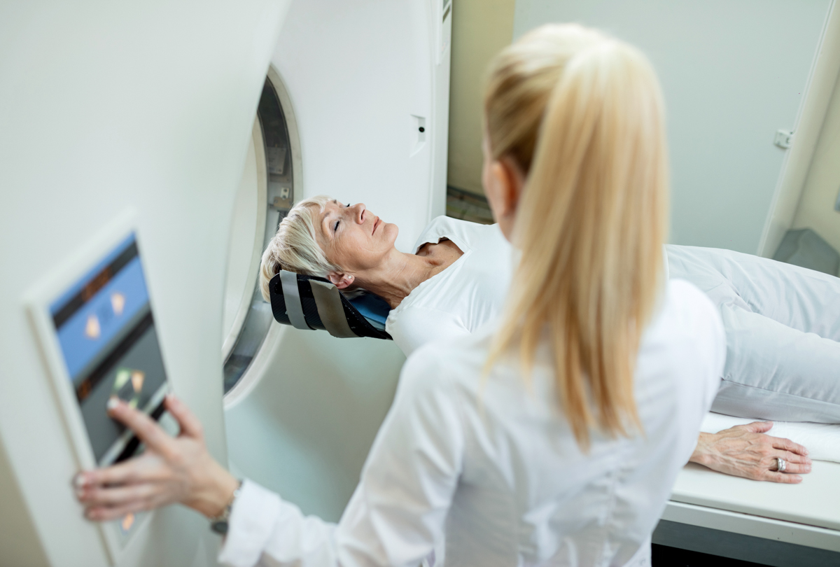
If you’ve been diagnosed with colorectal cancer, your care team will need to make sure that the cancer hasn’t spread. A computed tomography (CT) scan can determine if that’s happened. Metastasis (the spread of cancer) commonly happens in the liver. It can also happen in the lymph nodes and lungs.
This type of scan takes X-rays of your body from many angles. You lie on a table that moves into the center of a ring-shaped scanner, and a thin X-ray beam rotates around you. As the pictures are taken, a computer makes detailed images of the inside of your body. You might be given an injection of a contrast dye through an IV line into your vein. The dye helps any tumors show up clearly on the scans.
This scan takes a few minutes. You won’t feel the scan. But some people feel uncomfortable having to lie still on a narrow table during the test.
Magnetic resonance imaging
Magnetic resonance imaging (MRI) uses magnets, radio waves and a computer to take pictures of the inside of your body. This type of imaging does not use radiation, and dye may not be needed. This test may be used to help your care team learn about any lesions that showed up but were unclear during a computed tomography scan. It might be used to look for cancer in the liver, brain and spinal cord.
This imaging is not painful, but it can take a long time to complete. A scan may take up to an hour. During that time, you’ll need to lie flat and still on a table that’s moved into a long, narrow tube. If you don’t like tight spaces, this test might be stressful for you. Talk with your provider about any concerns.
The equipment makes loud banging noises during the scan. You can ask for earplugs if you think the noise will bother you.
Positron-emission tomography scan
A positron-emission tomography (PET) scan is a follow-up test that can help your care team understand whether an abnormal area seen on a different imaging test is cancer. It can also be used to determine if the cancer has spread to the lymph nodes or other parts of the body.
The picture isn’t as detailed as a computed tomography scan. But it can be used along with one done at the same time to look for tumors. This is called a PET-CT scan.
For this test, you are injected with a sugar containing a mildly radioactive substance. Cancer cells absorb more of this sugar than normal cells, and the radioactive material shows up during the image from the scan. During the scan, you’ll need to lie still on a table that slowly goes through the big rings of a positron-emission tomography scanner. This machine takes pictures that show where the sugar is in your body. The process may take a few hours. This type of scan is painless. But if you’re sensitive to the sugar, you may have nausea, headache or vomiting.
Ultrasound
Ultrasound uses sound waves to help your care team see the inside of your body. A small wand called a transducer is moved over your skin. It gives off sound waves. The computer picks up the echoes as they bounce off the body’s organs. Ultrasound can be used to look for tumors in your liver, gallbladder, pancreas or other places in your belly. But it can’t look for tumors of the colon.
For the exam, you lie on a table while the transducer is moved along the skin over the part of your body being checked. Usually, the skin is first lubricated with gel. The test is painless and fairly quick.
Two other kinds of ultrasound can be useful for colorectal cancer:
- Endorectal ultrasound. This can show how deep a tumor is in your rectum. It can also find lymph nodes that might be cancerous. This is important for finding out how advanced your cancer is. A thin probe is put into your rectum through a tool called a proctoscope. The full exam takes five to 10 minutes. It may be uncomfortable, but it isn’t painful. To get ready, you’ll need an enema the morning of the procedure. An enema involves inserting liquid directly into the rectum to help clean out the colon.
- Intraoperative ultrasound. For this, the probe is put against your liver during surgery. This test can help show if colorectal cancer has spread to your liver.
Chest X-ray
A chest X-ray can be used to see if the cancer has spread to your lungs. An X-ray uses radiation to make a picture of the inside of your body. The test is simple. It’s very good at showing lung nodules. It can also help show if you have lung or heart disease.
Working with your health care provider
Your health care provider will talk with you about which tests you’ll have. Make sure to get ready for the tests as instructed. Ask questions and talk about any concerns you have.